Houston Scoliosis and Spine Institute

Pedicle Subtraction Osteotomy
Patient History: 65-year-old male presented with complaints of back pain and leg weakness after a multilevel low back minimally invasive decompression. All conservative measures proved ineffective. His x-rays show collapse of lumbar spine into scoliosis. He was treated with a scoliosis correction and decompression. Approximately one year later, he noted an inability to stand upright and x-rays revealed a broken pelvic screw. He underwent a second procedure involving a complex wedge cut of the spine known as a pedicle subtraction osteotomy.
Surgery: First surgery involved passive realignment and scoliosis correction. Patient did well with this surgery until his pelvic screw broke. The second surgery involved a complex spinal maneuver known as the pedicle subtraction osteotomy. Additionally, a 3 rod construct was employed for additional stability and support.
Outcome: The patient now stands erect. He is able to walk further than before surgery. He still has mild back ache from time to time.
-
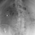
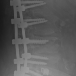
- Fig.1 AP View Lumbar Spine
-
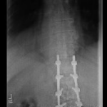
- Fig.1 PA ViewCorrected
-
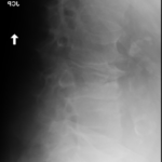
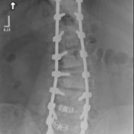
- Fig.2 Lateral View Lumbar Spine
-
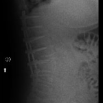
- Fig.2 Lateral ViewCorrected
Broken Iliac (Pelvic) Screw – Post op check up
-
- Fig.1 Frontal View Xray Showing Left Broken Iliac (Pelvic) Screw
-
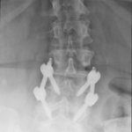
- Fig.2 Frontal View Xray Showing Re-Alignment after Wedge Osteotomy
-
- Fig.3 Side View Xray Showing Left Broken Iliac (Pelvic) Screw
-
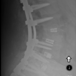
- Fig.4 Side View Xray Showing Re-Alignment After Wedge Osteotomy